
If you have had an ultrasound for a checkup or another reason and heard “your lining looks a bit thick,” that one remark can stay on your mind for days. Looking it up can be even more confusing, since every source seems to give a different number. Endometrial thickness is hard to understand as a number on its own; it can only be interpreted together with when and in what situation it was measured.
Earlier columns covered how to record bleeding between periods and the order in which the causes of abnormal bleeding are narrowed down. This time, we look separately at a finding you often hear along the way: a “thick lining.”
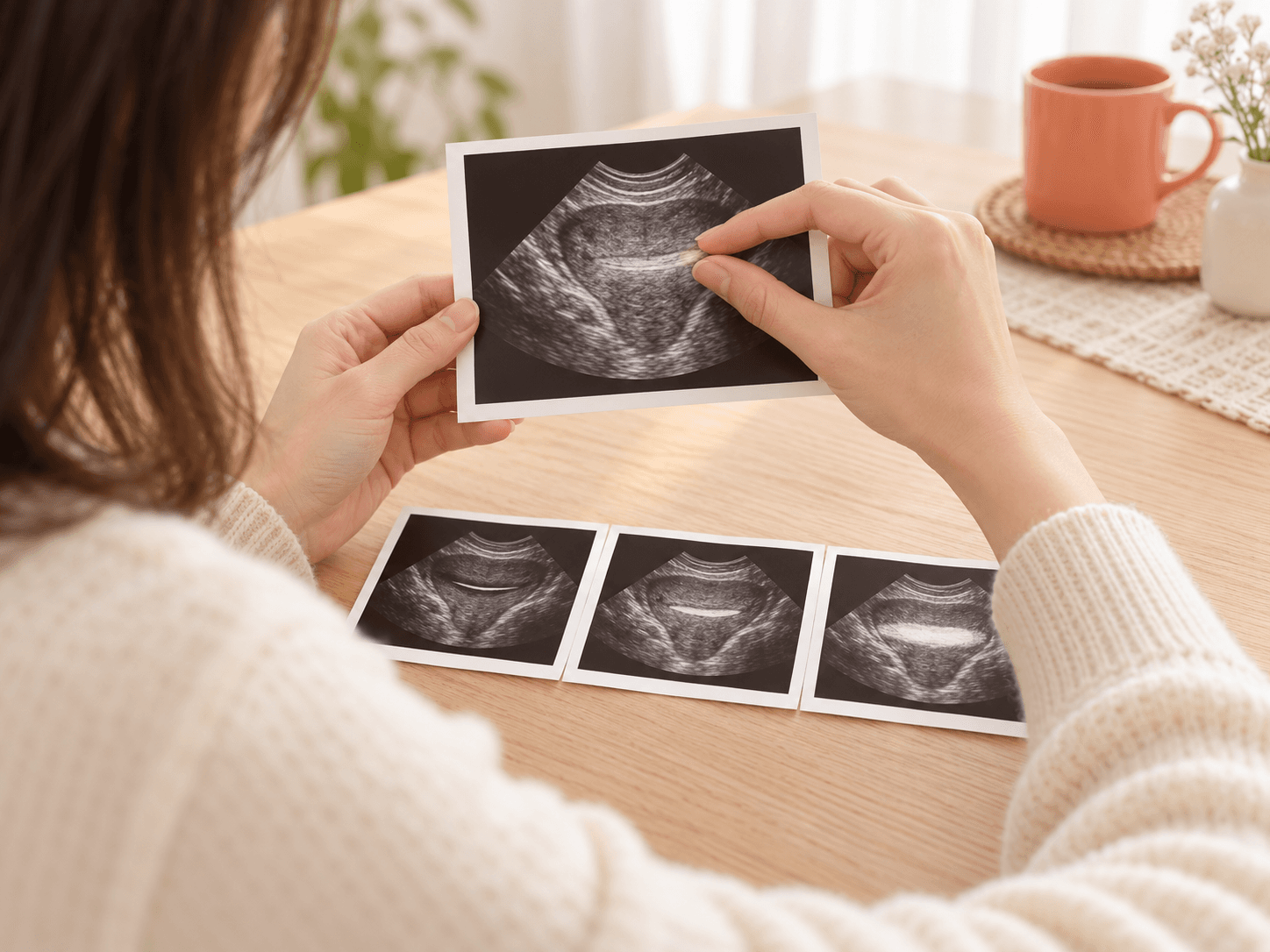
This generated image is for illustration and shows that the same uterus can have a different-looking lining depending on when it is viewed. It is not an actual test result.
Does “thick endometrium” mean something is wrong?
It is not a diagnosis in itself. It is an observation that the inner lining of the uterus measured thicker than expected on the ultrasound screen. It may be a natural change or a clue to a change that needs checking, so it is read together with other information.
The endometrium is the layer that covers the walls of the space inside the uterus, and it grows and breaks down over and over on a roughly monthly cycle. On the ultrasound screen it appears as a bright stripe running across the middle of the uterus, and the measured width of that stripe is the thickness on your report. To interpret this value, the following information is also needed.
- Date of the scan and the first day of your last period: This shows at what point in your cycle the measurement was taken.
- Whether you have gone through menopause: The standard used to read the value changes depending on whether 1 year has passed since your last period.
- Whether there has been bleeding: This includes bleeding between periods, periods that have become longer or heavier, and bleeding after menopause.
- Hormones and other medicines you take: Birth control pills, menopausal hormone therapy, and tamoxifen taken to lower the chance of breast cancer coming back can stimulate the lining.
- Shape of the lining: Whether it is evenly thick all over or one part bulges out is also a clue.
Why does endometrial thickness change from scan to scan?
Because before menopause, the endometrium is a tissue whose thickness can change several-fold within a single month. So even for the same person, the number can come out very differently depending on the day of the scan.
When a period ends, the lining starts again from a thin state. In the first half of the cycle it gradually thickens under the influence of estrogen from the ovaries, and after ovulation progesterone changes the lining into a state ready to receive a fertilized egg. If pregnancy does not occur, both hormones fall, and the lining sheds as a period.
One literature review summarizes that during a period the lining looks like a thin line of about 1~4 mm, and after ovulation it can measure up to 16~18 mm. Values vary between people and studies, but it is a natural pattern for the thickness measured just before a period to be much greater than just after one.
That is why, before menopause, a single rule such as “anything over a certain number of mm is abnormal” is not applied as is. If you were told your lining was thick around the time just before a period, without symptoms such as bleeding, you may be advised to have another look when the lining is thin, such as right after your period ends. When the lining is thin, it is easier to tell apart a change in which only one part stands out.
Before and after menopause, is the same thickness read differently?
Yes. After menopause the lining usually stays thin, so the same number is viewed more carefully. Above all, whether there has been bleeding after menopause is the most important dividing point.
When ovulation stops, the hormonal stimulation that makes the lining grow decreases, and the lining becomes thin. The Korean Society of Obstetrics and Gynecology describes the normal endometrial thickness in postmenopausal women not on hormone therapy as 5~6 mm or less, and as usually not exceeding 8 mm even during hormone therapy. During the menopausal transition, when cycles become erratic, it is hard to pinpoint the point in the cycle, so the bleeding pattern and age are given more weight.
| Situation | How the thickness is read | Checks that commonly follow |
|---|---|---|
| Before menopause, no symptoms | The normal range is wide depending on the point in the cycle, so no single cutoff is used | Repeat ultrasound when the lining is thin; further checks if the shape is uneven |
| Before menopause, with abnormal bleeding | The bleeding pattern, age, and risk factors are considered together, rather than thickness alone | Biopsy or hysteroscopy depending on age, risk factors, and response to treatment |
| After menopause, with bleeding | The American College of Obstetricians and Gynecologists considers endometrial cancer very unlikely if the lining is thin at 4 mm or less | Further checks such as a biopsy if over 4 mm; checked again if bleeding continues even at 4 mm or less |
| After menopause, no bleeding | Not everyone is tested just because it is over 4 mm | Decided individually based on risk factors such as age, obesity, and diabetes |
| On hormone therapy or tamoxifen | Medicines can make the lining look thicker, so it is interpreted separately | Unexpected bleeding is checked actively |
If you have no bleeding after menopause and happened to be told your lining is thick, the next plan is usually made by looking at your own risk factors rather than going straight to a biopsy on that basis alone. But if bleeding occurs after that, the assessment changes, so let your doctor know right away even if the amount is small.
What can make the lining thick?
The range is wide, starting with natural reasons such as the timing of the measurement and extending to endometrial polyps, endometrial hyperplasia, the effects of hormones and medicines, and, rarely, endometrial cancer. The ultrasound number alone cannot tell which of these applies, so further checks are needed.
- Timing of the measurement: If measured after ovulation or just before a period, even a normal lining comes out thick. This is the most common reason before menopause.
- Endometrial polyps: These are parts of the lining that grow out into the uterine cavity like small lumps, and most are benign. A polyp can make the whole lining look thicker. They are often found without any symptoms, but many are noticed because of bleeding between periods, and with bleeding after menopause or with older age, the uncommon chance that a cancerous change is also present becomes greater.
- Endometrial hyperplasia: This is a condition in which the cells of the lining have increased too much. It mainly develops when estrogen stimulation continues for a long time without enough progesterone. Without atypical cells, it can return to normal when the cause is managed, but with atypical cells the risk of progressing to cancer is higher, so the treatment approach is different.
- Effects of hormones and medicines: Polycystic ovary syndrome, in which ovulation is often skipped; obesity, in which estrogen is also made in fat tissue; hormone therapy using estrogen alone while you still have a uterus; and taking tamoxifen can all thicken the lining.
- Endometrial cancer: It is not a common cause, but it is a possibility that must be ruled out first when there is bleeding after menopause. Abnormal bleeding is the most common first sign.
Endometrial hyperplasia can only be confirmed by taking lining tissue and looking at it under a microscope. A thick lining on ultrasound is only a clue that “it could be hyperplasia,” not a sign that it is hyperplasia. The American College of Obstetricians and Gynecologists lists age 35 or older, never having been pregnant, an early first period or late menopause, obesity, diabetes, polycystic ovary syndrome, smoking, and a family history of uterine, colorectal, or ovarian cancer as risk factors linked to hyperplasia.
What is the next test, and how is it chosen?
If there is no bleeding and the timing of the measurement likely explains it, the ultrasound is repeated at a different time. If there is bleeding or risk factors, or if only one part stands out, a test that checks the lining directly is considered.
| Test | Considered when | Good to know |
|---|---|---|
| Repeat ultrasound | Before menopause, without symptoms, when the measurement was taken at a time the lining is thick, such as just before a period | It is repeated when the lining is thin, such as right after a period ends. Along with the number, it also looks at how the shape has changed. |
| Endometrial biopsy | When there is bleeding and the lining is thick overall, or when hyperplasia or cancer needs to be checked | Part of the lining is collected with a thin instrument. Because only part is obtained, a polyp in one spot can be missed, and the test may be repeated if the sample is not enough. |
| Hysteroscopy | When one part stands out, as with a polyp, or when bleeding continues even after a biopsy | A thin camera looks inside the uterus, and sampling the needed area or removing a polyp can be planned together. |
What to do first is decided by bringing together age and menopausal status, the pattern of bleeding, the shape seen on ultrasound, and previous results. Not everyone has both tests, and being recommended a test does not mean that cancer is suspected.
Both tests can cause pain like period cramps and bleeding, and rarely infection or injury to the uterus. If bleeding increases, abdominal pain becomes severe, or you develop a fever after the test, contact the facility that did the test right away. You can see the preparation and process in the endometrial biopsy guide and the hysteroscopy guide.
If a biopsy confirms hyperplasia, treatment depends on whether atypical cells are present. Without atypia, weight management is often combined with progestin medicines or a hormone device placed inside the uterus, followed by another biopsy after a set period. With atypia, treatment options including surgery are discussed. If a result calls for care at a higher-level hospital, please see the referral process described in the endometrial cancer / ovarian cancer information.
What symptoms need medical care right away?
Bleeding is a more important signal than the thickness number. Even while you are waiting for your next test date, do not put off getting care if you notice changes like those below.